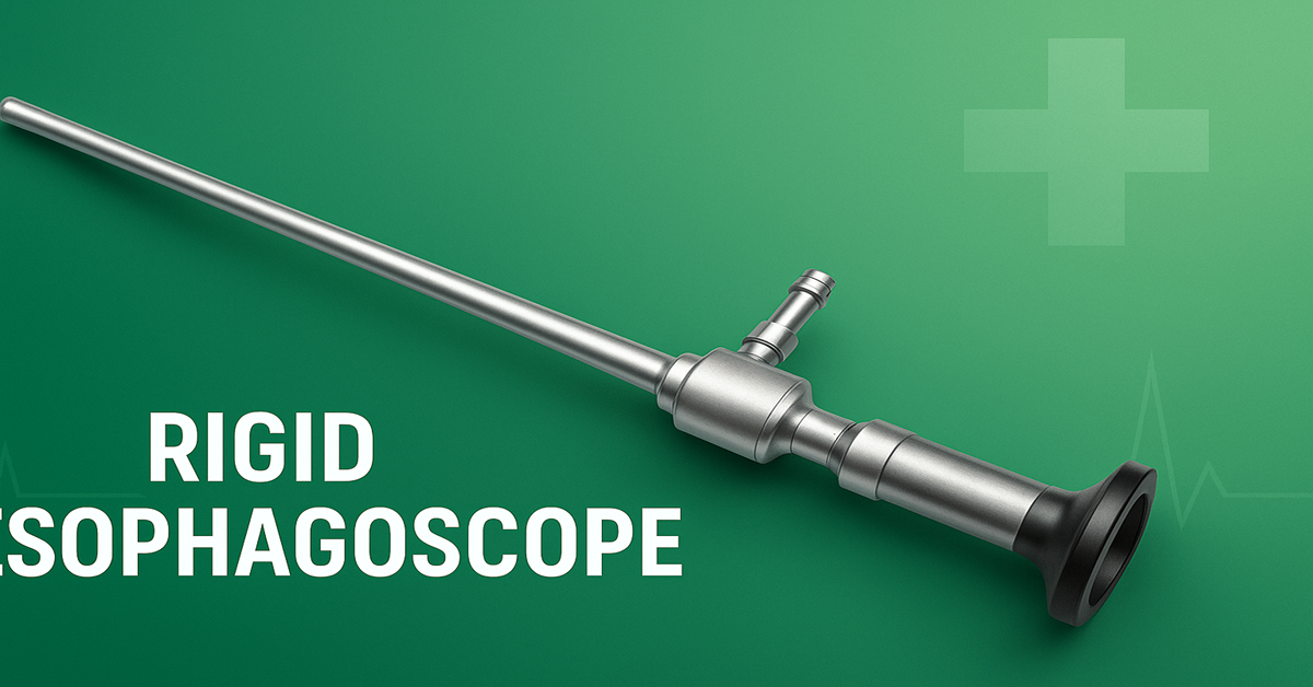
Rigid Esophagoscope: Advanced Uses, Challenges, and Clinical Relevance
The term Rigid Esophagoscope refers to a solid, non flexible instrument used to directly visualize, access, and intervene in the esophagus under direct line of sight control. In recent decades, video equipped rigid esophagoscopes have added the benefit of modern imaging to the traditional rigid technique. In this article, we explore the applications, advantages, limitations, procedural techniques, safety considerations, and evolving role of the rigid esophagoscope in contemporary medicine.
Introduction: The Place of Rigid Esophagoscope
Rigid esophagoscopy has a long and storied history in the domain of otolaryngology and upper gastroenterology. Though many centers now favor flexible endoscopes, rigid esophagoscopes especially those with video capability continue to play a vital role in certain diagnostic and therapeutic settings. The rigid esophagoscope provides unmatched stability, direct instrument control, and the ability to manipulate foreign bodies or strictures under direct vision with minimal ambiguity. The integration of video capability into rigid scopes combines the strengths of rigid mechanics with modern imaging, making them valuable tools in the hands of experienced clinicians.
The rest of this article will examine how rigid esophagoscopy is used, what its benefits and risks are, how procedures are performed, and how its role is evolving.
Historical and Technological Context
Rigid esophagoscopes have been used for over a century to access the esophagus directly. Early instruments were simple metal tubes with basic illumination and limited optics. Over time, rod-lens systems improved visualization, and later, miniaturized cameras and digital video modules have been incorporated into rigid designs, giving rise to video rigid esophagoscopes. These hybrids allow the clinician to benefit from high-definition imaging while retaining the mechanical advantages of rigidity.
The video rigid esophagoscope is designed so that the clinician can both see the internal lumen and manipulate instruments through accessory ports, allowing biopsy, foreign body removal, dilation, or hemostasis under direct view.
Clinical Indications of Rigid Esophagoscopy
Rigid esophagoscopy remains especially useful in several scenarios:
Foreign Body Retrieval
One of the most common and compelling indications is the removal of ingested foreign bodies that have become lodged in the esophagus. Sharp objects, coins, dentures, fish bones, or other items can be grasped more securely under direct rigid control. Studies report high success rates for rigid instrument retrieval (often 94 100 %) with low incidence of complications such as perforation when performed by skilled operators.
Stricture Assessment and Dilation
Rigid scopes allow controlled inspection and management of strictures, especially in the upper or cervical esophagus. Under direct visualization, the clinician can safely dilate or incise scar tissue while monitoring mucosal integrity.
Biopsy and Lesion Evaluation
Suspicious mucosal lesions, superficial tumors, or irregularities can be biopsied under direct view. The strength and direct access of rigid scopes help with precise sampling.
Hemostatic and Therapeutic Intervention
Bleeding lesions, small tumors, or localized pathology may be managed via electrocautery, clipping, or cutting under rigid scope guidance. Accessory tools can be passed reliably, and the treatment can be more controlled.
Training, Teaching, and Resource‑Limited Settings
Rigid techniques are often taught in ENT and surgical training programs to help students understand direct-line anatomy and instrument behavior. In low-resource or rural settings where flexible instruments might not be available or maintained, rigid esophagoscopy provides a robust alternative with lower maintenance demands.
Advantages of Rigid Over Flexible Techniques
Rigid esophagoscopy continues to offer certain advantages even though flexible endoscopes dominate many settings:
-
Superior Stability and Tactile Feedback
Because the rigid tube does not bend, forces are transmitted more predictably. The operator can feel resistance, torque, or engagement, which is useful when manipulating tight strictures or removing foreign bodies. -
Better Instrument Control
Accessory instruments (forceps, graspers, dilators) behave more predictably, with less risk of bending or buckling inside the working channel. -
High Optical Clarity
The optics of rigid systems often deliver sharper, distortion‑free images, especially when paired with modern video chips. -
Efficient Foreign Body Management
For large, sharp, or irregular objects, the directness of the rigid path helps reduce manipulation time and risk. -
Durability and Sterilization Resilience
Rigid scopes have fewer delicate parts; they tolerate repeated sterilization cycles more robustly than fiberoptic or flexible scopes. -
Lower Repair Overhead
Because they have simpler mechanical and optical components, rigid systems may require fewer repairs or adjustments over time.
Limitations and Challenges
The use of rigid esophagoscopes also comes with significant challenges and risks:
-
Patient Discomfort and Need for Anesthesia
Rigid instruments are more invasive, and many procedures require general anesthesia or deep sedation to avoid gag reflex, pain, or trauma. -
Anatomic Constraints
Because of rigidity, the device cannot negotiate sharp curves or tortuous anatomy. Lesions in the distal esophagus or near the gastroesophageal junction may be out of reach. -
Risk of Perforation and Trauma
Inexperienced users or pathologic conditions (e.g., fragile mucosa) may result in tears or perforations. Esophageal perforation, while uncommon, is a serious complication. -
Blind Spots
Certain anatomical niches might not be visualized if the rigid path cannot align perfectly. -
Steep Learning Curve
Many clinicians today are more experienced with flexible endoscopy, and mastering rigid techniques requires deliberate practice. -
Limited Flexibility in Complex Cases
For very distal or tortuous disease, rigid tools may not suffice.
Procedural Technique and Best Practices
To maximize success and minimize risk, rigid esophagoscopy must be conducted carefully:
-
Preoperative Planning
Study imaging (X-rays, CT, barium studies) to locate the lesion or foreign body and anticipate anatomical challenges (e.g., cervical spine disease). -
Anesthesia and Airway Management
General anesthesia with endotracheal intubation is common for rigid esophagoscopy. In selected cases, deep sedation may suffice if patient cooperation is adequate. -
Patient Positioning
Typically supine, with neck extension aligned straight with the thoracic spine. Proper head support is essential, particularly in elderly patients with limited cervical mobility. -
Gentle Introduction
The rigid tube is advanced carefully, often along the posterior pharynx, avoiding undue pressure on sensitive structures. -
Insufflation, Irrigation, and Suction
Light insufflation may help distend the lumen; irrigation and suction are used to clear secretions and maintain visibility. -
Systematic Inspection
Begin proximally and move distally, noting mucosal patterns, strictures, anomalies, or foreign objects. -
Safe Instrument Manipulation
Use appropriate forceps, snares, or retractors under direct vision. Do not attempt blind retrieval. -
Hemostasis and Assessment
Control bleeding with irrigation, pressure, or cautery. Before withdrawal, inspect the mucosa for injuries or perforation signs. -
Postprocedural Monitoring
Watch for signs of perforation (pain, crepitus), bleeding, or infection. Imaging (contrast esophagram, CT) may be warranted if complications are suspected. -
Documentation
Record all findings, procedure steps, accessory tools used, video or still images, and any complications.
Safety, Risks, and Complication Management
Rigid esophagoscopy carries risks that must be anticipated and mitigated:
-
Perforation / Esophageal Tear
Although uncommon, perforation is a serious complication. Operators must be willing to abort a difficult passage and refer or convert to alternative techniques. -
Bleeding
Vascular lesions, biopsies, or inadvertent mucosal trauma may cause bleeding, which must be managed promptly. -
Mediastinitis / Infection
A perforation may lead to leakage and mediastinal infection, which can become life-threatening. -
Airway Complications
Because the procedure occurs through the upper airway, vigilance for aspiration, obstructed tube, or airway injury is crucial. -
Dental / Pharyngeal Trauma
Teeth, soft palate, or oropharyngeal tissues may be damaged during insertion if protective measures (e.g., tooth guards) are not used.
To manage these risks, clinicians should:
-
Work within their skill set and refer when anatomy or pathology is complex.
-
Use gentle technique and avoid forcing passage.
-
Monitor vital signs and oxygenation continuously.
-
Have readiness for emergency conversion, perforation repair, or surgical backup.
-
Practice in simulation or cadaver settings to maintain competence.
Rigid Versus Flexible Esophagoscopy: Complementary Tools
While flexible endoscopy is now the standard for many esophageal evaluations, rigid and flexible techniques often complement each other:
-
Reach
Flexible scopes can navigate long, tortuous, or distal paths; rigid scopes excel in straight-line, proximal access. -
Comfort
Flexible scopes may be done under sedation; rigid ones often need general anesthesia. -
Instrumentation Control
Rigid scopes offer more direct, robust control over devices; flexible channels may limit instrument rigidity. -
Foreign Body Management
Rigid scopes are preferred for large, sharp, or impacted objects; flexible scopes may suffice for small, soft, or distal ones. PubMed+2PMC+2 -
Diagnostic Versatility
Flexible scopes can examine stomach, duodenum, or perform extended biopsies; rigid scopes are more focused on the esophagus.
Thus, a thoughtful clinician will choose the modality best suited to the case, sometimes using both in tandem.
Innovations and the Future of Rigid Esophagoscope
Rigid esophagoscopy is advancing to meet modern expectations. Some emerging trends and innovations include:
-
Video Integration
Higher resolution CMOS or CCD sensors are increasingly embedded in rigid scopes, offering real time imaging and digital documentation. -
Modular Design
Interchangeable heads, optics, and accessory ports highlight flexibility without sacrificing rigidity. -
Angulated Rigid Tips
Some rigid scopes now offer limited articulation near the tip to negotiate minor curvatures without losing stability. -
Hybrid Devices
Combining a rigid external frame with a flexible insertion tip may offer the best of both worlds in select applications. -
Tele‑assistance and Training
Live remote guidance during rigid procedures could expand use in underserved areas. -
Miniaturization and Material Advances
Stronger but lighter materials may reduce the diameter of rigid scopes while maintaining structural integrity.
As these technologies mature, rigid esophagoscopy may take on new roles and reach broader acceptance in challenging clinical settings.
Case Illustrations
Case 1: Fish Bone Impaction
A middle-aged patient presents with throat pain and dysphagia after ingesting fish. Imaging suggests a sharp bone lodged at the upper esophagus. Under general anesthesia, a video rigid esophagoscope is introduced; the bone is visualized and grasped securely with forceps, then removed with minimal mucosal trauma demonstrating how rigid control and direct imaging expedite safe extraction.
Case 2: Tight Proximal Stricture
A patient with progressive dysphagia is discovered to have a ring-like stricture just distal to the cricopharyngeus. Using rigid esophagoscopy, the clinician advances to the stricture, visualizes it directly, and performs cautious incision and dilation under view, monitoring for mucosal integrity. The procedure restores lumen patency without perforation.
Case 3: Mucosal Lesion Biopsy
A patient with intermittent odynophagia is found to have a suspicious mucosal irregularity in the upper third of the esophagus on imaging. The rigid scope is used to evaluate the lesion; targeted biopsies are obtained under clear view, demonstrating precise sampling with minimal risk. Histopathology reveals early dysplasia, allowing timely intervention.
Training, Competency, and Clinical Adoption
Given the specialized nature of rigid esophagoscopy, maintaining proficiency is critical. Key strategies include:
-
Structured Training Programs
Trainees should begin with simulation, followed by supervised clinical cases. Cadaver labs or bench models help in mastering instrument control and safe technique. -
Regular Case Exposure
Because rigid methods are less commonly used than flexible ones in many centers, deliberate scheduling of cases ensures skills do not atrophy. -
Complication Drills
Simulation of perforation, bleeding, or airway emergencies fosters readiness. -
Interdisciplinary Collaboration
ENT, GI, thoracic surgery, and anesthesiology coordination helps institutionalize rigid modality use. -
Research and Audits
Collecting outcome data, complication rates, and procedural success benchmarks fosters continuous improvement and justification of rigid esophagoscope use.
Conclusion
The Rigid Esophagoscope, particularly in its modern video‑enhanced form, remains an important and versatile tool in esophageal diagnostics and intervention. While flexible endoscopy may dominate routine evaluations, rigid technology offers unique advantages in foreign body retrieval, stricture treatment, lesion biopsy, and controlled instrumentation. Its durability, optical clarity, and mechanical predictability make it indispensable in many contexts, especially when handling challenging or high risk interventions.
Effective use of rigid esophagoscopy requires experienced hands, careful planning, and attention to safety. With continued innovation such as better imaging, modular designs, and hybrid systems the rigid esophagoscope is likely to retain a meaningful role in future clinical practice. Rather than being supplanted, it is evolving to meet 21st century needs, and remains a critical component in the armamentarium of skilled endoscopic practitioners.
If you like, I can also provide a version tailored to non specialists or a summary you can use for hospital websites or patient education. Do you want me to adapt it further?